Ultrasound Scans
What is an Ultrasound Scan?
Ultrasound, also known as sonography, is a non-invasive diagnostic imaging technique that uses high-frequency sound waves to create real-time images of the body’s internal structures. It is widely used in medicine due to its safety, effectiveness, and versatility.
Unlike X-rays or CT scans, ultrasounds do not use ionizing radiation, making them especially safe for sensitive populations like pregnant women and children.

How Does an Ultrasound Work?
The basic principle of ultrasound imaging involves sound waves and their echoes:
Sound Wave Transmission:
- A handheld device called a transducer emits high-frequency sound waves into the body.
Echo Formation:
- These sound waves bounce off internal structures, such as organs, tissues, and fluids, creating echoes.
Image Generation:
- The machine processes these echoes into visual images, displayed on a monitor in real-time.
This process is entirely painless and typically takes less than 30 minutes.

Types of Ultrasound Scans
- Abdominal Ultrasound: Examines organs in the abdominal cavity, such as the liver, kidneys, and gallbladder.
- Pelvic Ultrasound: Focuses on the reproductive organs in women and the bladder in men.
- Obstetric Ultrasound: Monitors pregnancy, fetal development, and potential complications.
- Musculoskeletal Ultrasound: Detects soft tissue injuries, such as ligament tears or muscle damage.
- Doppler Ultrasound: Evaluates blood flow in veins and arteries, often used to detect blockages or clots.

Key Applications of Ultrasound
Ultrasounds are used in various medical scenarios, providing critical information for diagnosis and treatment:
Pregnancy:
- Ultrasound is indispensable during pregnancy to:
- Confirm fetal heartbeat and monitor development.
- Detect abnormalities, such as ectopic pregnancy or birth defects.
- Estimate due dates and assess amniotic fluid levels.
- Ultrasound is indispensable during pregnancy to:
Gallstones:
- Gallstones are solid deposits in the gallbladder, often diagnosed using abdominal ultrasound. This test helps identify:
- Size and number of gallstones.
- Inflammation or blockages in the bile ducts.
- Gallstones are solid deposits in the gallbladder, often diagnosed using abdominal ultrasound. This test helps identify:
Liver Issues:
- Ultrasound effectively evaluates liver conditions, such as:
- Fatty liver disease.
- Liver cirrhosis.
- Tumors or cysts.
- Ultrasound effectively evaluates liver conditions, such as:
Kidney Stones:
- Kidney stones are mineral deposits that form in the kidneys, causing severe pain. Ultrasound helps:
- Locate stones and measure their size.
- Assess damage to kidney tissue.
- Kidney stones are mineral deposits that form in the kidneys, causing severe pain. Ultrasound helps:
Soft Tissue Injuries:
- Ultrasound is a go-to diagnostic tool for sports injuries, including:
- Torn ligaments.
- Muscle tears or strains.
- Fluid buildup in joints.
- Ultrasound is a go-to diagnostic tool for sports injuries, including:
The Ultrasound Procedure
Preparation:
- In most cases, no special preparation is required. However:
- For abdominal ultrasounds, fasting for 6–8 hours may be recommended.
- For pelvic scans, a full bladder is often required to improve visibility.
- In most cases, no special preparation is required. However:
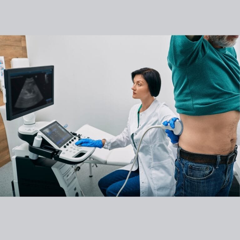
Scanning Process:
- The patient lies down, and a clear gel is applied to the skin to enhance sound wave transmission.
- The technician moves the transducer over the targeted area to capture images.
Duration:
- The procedure is quick, typically lasting 15–30 minutes.
Post-Scan:
- Results are interpreted by a radiologist and shared with the referring physician.
Advantages of Ultrasound
- Non-Invasive and Safe: No exposure to radiation.
- Real-Time Imaging: Allows immediate evaluation of moving structures like the heart or fetus.
- Wide Range of Uses: Applicable for diagnostics, monitoring, and guiding procedures like biopsies.
- Cost-Effective: Generally less expensive than other imaging modalities like MRI or CT scans.
Limitations of Ultrasound
- Limited Penetration: Cannot image deep tissues or bones effectively.
- Operator Dependence: Image quality relies heavily on the skill of the technician.
- Low Sensitivity for Certain Conditions: May not detect very small tumors or subtle abnormalities.
FAQs About Ultrasound Scans
1. Who Can Perform an Ultrasound?
Ultrasound scans are conducted by trained sonographers or radiologists who are skilled in using the equipment and interpreting images.
2. What Qualifications are Needed to Operate an Ultrasound Machine?
- A degree or diploma in Medical Imaging Technology or Radiology.
- Specialized training in ultrasound technology.
- Certification from recognized boards or councils.
3. Are There Any Risks Associated with Ultrasound?
Ultrasound is considered extremely safe with no known risks when performed by qualified professionals.
4. Can Ultrasound Be Performed During Pregnancy?
Yes, ultrasound is commonly used during pregnancy and is completely safe for both the mother and fetus.
5. How Much Does an Ultrasound Cost?
The cost of ultrasound varies depending on the type and healthcare facility. On average:
- Abdominal Ultrasound: ₹1,000–₹2,500.
- Pregnancy Ultrasound: ₹800–₹2,000.
- Doppler Ultrasound: ₹2,000–₹5,000.
6. What is the Process for Setting Up an Ultrasound Lab?
- Licensing and Approvals:
- Obtain necessary permissions from health regulatory bodies in your country.
- Equipment:
- Invest in a reliable ultrasound machine, costing ₹5 lakhs to ₹25 lakhs.
- Infrastructure:
- Minimum space of 200–300 square feet is required for the lab.
- Staffing:
- Hire qualified sonographers, technicians, and administrative staff.
- Compliance:
- Ensure the lab meets safety and hygiene standards.
Conclusion
Ultrasound scans are an essential diagnostic tool, offering safe, non-invasive, and real-time imaging for a variety of medical conditions. Whether monitoring a pregnancy, diagnosing gallstones, or evaluating soft tissue injuries, ultrasound technology continues to play a pivotal role in modern healthcare. With its affordability, versatility, and safety, ultrasound remains a cornerstone of diagnostic medicine.